Plant Tissues
Plants that we observe around us are
usually multi-cellular. They perform
several life processes such as growth,
respiration, excretion, etc, similar to those
performed by animals. In addition to these
they can perform photosynthesis and
prepare food not only for themselves but
also for all the other living organisms
dependent on them, either directly or
indirectly.
Let us recall the information about
different parts of the plants and the
functions they are associated with.
Activity-1:
Parts of the plant and their
functions.
We have studied about the functions of
the parts of the plant in the earlier classes.
Read the functions given below and write
the names of the parts involved in
performing the respective function.
Function Name of the parts:
1. Absorption of water from soil
2. Exchange of gases (air)
3. Photosynthesis
4. Transpiration
5. Reproduction
Activity-2:
Cells in onion peel
- Take a piece of an onion peel.
- Now place it on the slide.
- Put a drop of water and then a drop of
glycerine on it.

- Gently cover it with a cover -slip.
- Observe it under the microscope.
- Draw and label the diagram that you
have observed under the microscope.
Compare your drawing with figure-1 to
find out labelled parts.
Are all the cells similar?
How are the cells arranged?
Activity-3:
Cells in a leaf peel.
- Take a betel leaf or Tradescantia or
Rheo leaf.
- Tear it with a single stroke. So that a
thin edge be seen at the torn end.
- Observe the thin edge where the leaf

has been torn under the microscope
in the same manner as you had
observed the onion peel.
Draw a diagram of what you have
observed and compare it with figure-2.
Are all the cells similar?
Is there any difference in their
arrangement?
What can we infer from the above
activities?
Have you noticed that the cells are in
groups in both the activities?
Compare and write a note on the
arrangements of the cells that you have
observed in both of the activities.
You may have observed that the cells are
present in groups with certain
arrangement. With the help of the
following activities we shall try to find out
whether these arrangements have special
roles to play in the plant body.
Activity-4:
(a) Cells in root tip
- Are the cells in the root similar to that
of a leaf. Let us find out how the cells
are arranged in the root. For this we
need onion root tips.
- Take a transparent (plastic/glass) bottle
filled with water. Take an onion bulb
slightly larger than the mouth of the
bottle.
- Put the onion bulb on the mouth of the
bottle.

- Observe the growth of roots for a few
days till they grow to nearly an inch.
- Take the onion out and cut some of the
root tips.
- Take an onion root tip.
- Place it on the slide.
- Put a drop of water and then a drop of
glycerine on it.
- Cover it with a cover-slip.
- Remove excess water with the help of
blotting paper.
- Tap the cover-slip gently and press with
the blunt end of the needle or brush to
spread the material.
- Observe the structure and arrangement
of the cells.
- Draw the diagram of that you have
observed under the microscope.
· Are all the cells similar?
· How is the arrangement of cells?
Cells in Onion roots tip:

Activity-5:
(b) Growing roots
- Take the onion used in the previous
activity and mark the cut end of the
roots with a permanent marker.
- Put the onion bulb on the mouth of the
bottle in the same way as you did in the
previous activity.
- Leave the set up aside for at least four
to five days. Take care that there is
enough water in the glass so that the
roots are submerged.
Did all the roots grow in a same manner?
What happened to the roots which had
been cut off?
Write down your findings, regarding the
cut roots and those that were not?
We observe that by removing the tip of
the onion root, having a particular
arrangement of cells, the growth of the root
in length is stopped.
You have observed that cells are present
in groups. Cells in groups which are nearly
similar in structure perform similar
functions. Such groups of cells are called tissues.

Growth on roots in onion bulb
Types of plant tissues:
There are four basic types of tissues in
the plants. They are
Tissues that bring about overall growth
and repair are called Meristematic
tissues.
Tissues that form outer coverings are
called as Dermal tissues.
Tissues that form the bulk of the plant
body, helping in packing other tissues
are called as Ground tissues.
Tissues that help in transport of
materials are called as Vascular tissues.
You have already observed some types
of tissues. To observe various types of plant
tissues we need to know some techniques
for preparing slides and cutting sections.
See annexure for the same.
1. Meristematic tissues:
Observe the given figure of a stained
section of a shoot tip

From the above figure you can infer
that meristematic tissues are present on
the tip, sides and in between layers of other
tissues. Meristematic tissues at the
growing tip that bring about growth in
length are called as apical meristematic
tissues.
Tissues present around the edges in a
lateral manner and giving rise to growth in
the girth of stem are called lateral
meristematic tissues.
Areas from which branching takes place
or a leaf or a flower stalk grows, we find
a kind of meristematic tissue called
intercalary meristematic (also called
Cambium) tissue.
Activity-6:
Dicot Stem tissues
Prepare a temporary mount of the TS
of a dicot stem.
Observe it under the microscope.
Draw and label the diagram.
Compare it with figure given below
Dicot stem (TS) - Tissues

What are the similarities between the
tissues in root and in shoot as shown
in the above figure?
Are all the cells similar in shape and
structure?
Activity-7:
Rheo leaf - Dermal tissue.
- Take a fresh leaf of Rheo or Betel plant
- Tear it in a single stroke, so that a thin
whitish edge can be seen at the torn
end.
Slowly remove it and observe that peel
under the microscope (by preparing a
temporary mount).
See the arrangement of cells .Are all
cells similar? Are there any spaces
between the cells?
This activity shows a part of the dermal
tissue of the plants.
Dermal tissue (Dermis) usually consists
of a single layer of cells showing variations
in the types of cells on the basis of their
functions and location. These tissues are
divided into three different types. They are
epidermis (outer most layer), mesodermis
(The middle layer) and endodermis (the
innermost layer).
The walls of the cells of dermal tissue
are comparatively thicker than the cells of
meristematic tissues. In desert plants it may
be even more thick and waxy. Small pores
are seen in the epidermis of the leaf, called
stomata. They are enclosed by two kidney
shaped cells, called guard cells. Cells of
the roots have long-hair like parts, called
root hair.

Parenchyma:
The cells of the parenchyma are soft,
thin walled and loosely packed. The
Parenchyma which contains chloroplasts is
called “Chlorenchyma”. The Parenchyma
which contains large air cavities or spaces
is called “Aerenchyma”. The Parenchyma
which stores water or food or waste
products is called “Storage Tissue”.
Collenchyma tissues have thick walled,
longer cells when compared with parenchyma.
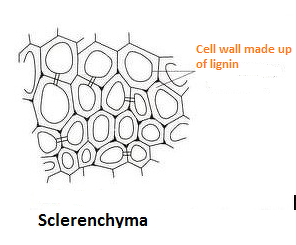
In the sclerenchyma the cells are thick
walled and tightly packed without spaces.
So when compared with dermal tissue, the
ground tissue is of various types. And some
of the ground tissues help in
photosynthesis.
Let us observe the ground tissue of
some other stems.

Activity-8:
Take permanent slides of Chlorenchyma, Arenchyma, Storage Tissue from your laboratory and observe them under the microscope. Find out the characteristics differences and record them in your notebook.
Chlorenchyma

Arenchyma

Storage Tissue

Vascular Tissue:
We know that roots can absorb water
from the soil and send it to other parts of
the plant. The leaves and other green parts
prepare food and supply it to all the parts
of the plant.
Let us study the tissues involved in
transportation.
We have done an experiment on
transportation in class VII, in the chapter
on plant nutrition. We have observed that
when a plant is kept in red coloured water,
Some of the parts of the plant turned red.
Do the same experiment again by keeping
a small plant (with roots) in red coloured
water. Leave it for two hours. Now cut a
T.S. of the stem and observe it under the
microscope.
Which part of the plant is responsible
for this transport?
Draw a rough sketch of that part and
mark the part that appears red.
What do you conclude from your observation?
The tissues involved in transportation are vascular tissues. They are composed of different
types of cells which show specific arrangements.

No comments:
Post a Comment