Tuesday, January 18, 2022

BIO(Diversity)

    Diversity in Living Organisms

        
      There are many plants and animals
around us. We know very little about them.
Most of them belong to a world not visible
to the naked eye, as you have already studied
in the chapter on, ‘Microbial World’. The
types of organisms that we have studied so
far are also in lakhs, existing from mountain
peaks to deserts and plateaus to the deep
oceans, from extreme cold conditions to
extreme hot ones and many more, such
diversity is the symbol of nature.
Studying about diversity as it is, would
be a very chaotic and difficult task.
Moreover describing and naming each
organism individually without knowing the
organisms that might be sharing common
characteristics would be insignificant. Thus
people who have tried to study diverse
organisms in nature have tried to make
groups of them on the basis of differences
and similarities found among them. This
helped to identify largely varied and closely
related groups of organisms.
Thus our knowledge of the entire living
world depends on first making meaningful.
groups to carry out our study in a
systematic manner.
In this lesson we will try to study the
diversity present among several living
organisms, classify and appreciate nature's
miracle

Diversity in plants:
Here as we finish our activity we would have established some common characteristics of land plants- those having two seed leaves are called dicotyledons, while those having single seed leaf are
called monocotyledons.


They share some common
characteristics like venation (dicots have
reticulate/branched, while monocots have
parallel venation).
By doing the above activity we can
understand how grouping is done in biology
by observing the similarities and
differences among diverse groups in the
sample under study. We will do some
similar exercises with animals now.

Diversity in animals:
Observation of external characters of insects Collect housefly, mosquito, ant, dung beetle, butterfly, moth and cockroach from your surroundings. Observe them carefully. Take the help of a magnifying lens to get a closer view. Are all insects of the same size or shape?

What differences did you observe
with regard to legs?
What differences did you observe
with regard to wings?
Is there any relationship between
the number of wings and legs?
Did you find any two insects with same
characters? If yes, display in the class. If
no, note down the differences in your note
book.
Even though all these are insects and
you see that they show several differences.
Can you find at least one character that is
similar to the whole group, what is it?
How do you group insects? Would it
be based on number of body segments or
number of legs they have?

The examples of insects given above
are of different species. Hence they show
a lot of difference and we say they are
diverse. If we were to compare insects of
the same type that is to say two houseflies
we will be perhaps still find some
differences(try it out yourself) and these
would be variations.
Let us see some variations that are
present in human populations

Monera
Observe the given slides carefully and
say
How many cells are found in the
organism?
Do you find any nucleus in the
middle of the cell?
Are there any other cell organelles
found in the cell?
By observing the above characteristics
we conclude that Monerans are:
A. One-celled organisms
B. Cells have no membrane bound nucleus
C. Reproduce by splitting into two.
D. Absorb nutrients from outside their
bodies
E. They move with the help of
locomotory organs like flagella, cilia
or hair like structures present on them.
F. Some monerans cause diseases, but
others are helpful to people.
G. Examples: bacteria

        Nostoc


        Bacteria


Three major groups of organisms come under this group. They are archaebacteria (ancient bacteria present till date, some species found in hot springs come under this), eubacteria (streptococcus, rhizobium, e.coli etc) and cyanobacteria which are also called blue green bacteria as they appear similar to blue green algae externally but internally are more like bacteria(but they are not bacteria).


Protista (protoctista):
Observe the given slides carefully and
say
How many cells are found in the
organism?
Do you find any nucleus in the
middle of the cell?
Are there any other cell organelles
found in the cell?
Are there any locomotory organs
in them?
Characteristics of protists
A. Most are one-celled (unicellular),
but some have many cells.
B. Cells have a membrane around the
nucleus.
C. Some get nutrients and energy by
eating other organisms.
D. Some get energy from the sun, and
nutrients from the water around
them.
E. These live either solitary or in a
colony.
F. Some of the cell organelles are
present inside the cell.
G. Mostly reproduce by splitting into
two (Binary fission).
H. Examples are paramoecium,
amoeba, algae, etc.

              Amoeba


           Paramecium


Fungi:
Observe the specimen and diagrams
given below and answer the following
questions.
What is the colour? Can they
prepare their own food as green
plants?
Make a sketch of the main parts of the
body
Do you find root like structures?
Guess why?

Characteristics of fungi
A. Most are many-celled (multi
cellular) and some are one-celled
organisms.
B. Eukaryotes with well defined
prominent head (you usually see
them propping out from the ground
or on barks of trees during rainy
season).
C. Get nutrients and energy by
absorbing/ digesting the surface
they live on through root like
structures which are fine thread
like parts of their body.
D. Most of these reproduce by spores.
E. Examples are yeast, mushrooms,
bread moulds, and lichens.


Plantae:
Several plants grow around you. Do
all of them produce seeds?
Think if grass produces seeds
(hint:compare with rice plants and
think).
Name some plants that produce
seeds.
Which part of the plant produces
seeds? Where is it located?(recall
structure of plant parts studied in
earlier classes)
Do all plants have a definite
structure to produce seeds?
Plants are diverse in nature. The basis
of classifying them is the way they acquire
their food, the type of reproductive
structures they have and the way they
reproduce. They are multicellular,
eukaryotic with cell walls. They are usually
autotrophs and use mainly chlorophyll for
photosynthesis.
The first level of classification among
plants depends on whether the plant body
has well differentiated, distinct parts.
The next level of classification is based
on whether the differentiated plant body has
special tissues (vascular tissues) for the
transport of water and other substances
within it. Further classification looks at the
ability to bear seeds and whether the seeds
are enclosed within fruits.
Let's look at some plants like moss and
ferns more closely.

            Funaria


Activity:
Observation of moss plants through
hand lens.
You can collect mosses from the
greenish velvety growth on bricks during
the rainy season. Scrap a bit of this greenish
growth over a slide and observe with a hand
lens or under a dissection microscope. 
These are not exactly flowers but
structures that contain seed like structures
called spores. Spores contain very little
food while the seed stores a lot of it.
Moreover where seeds are produced from
ovule of flower, spores are produced within
structures called as sporangium in a
different manner.

 Sporophylls of Cycas & Fern


Coelenterata/Cnidarians:

These are aquatic forms showing more body design differentiation when compared with Poriferans. There is gastrovascular cavity in the body. The body is made up of two layers of cells: one forming the outer layers while the other forming the inner layers. Some
live in colonies ,like the corals that are tiny nearly 3 to 56 mm but their colonies where we may find several types of them which
form coral islands huge as say an island (1800 sqkm ), is called coral reefs while others like hydra, jellyfish and sea anemones are common examples.

Hydra


Platyhelminthes:
The body of animals in this group is far
more complexly designed than in the two
other groups we have considered so far. The
body is bilaterally symmetrical, meaning
that the left and the right halves of the body
have the same design. There are three layers
of cells from which differentiated tissues
can be made, that is why such animals are
called triploblastic. This allows outside and
inside body linings as well as some organs
to be made. There is thus some degree of
tissue formation. However, there is no true
internal body cavity or coelom, in which
well developed organs can be
accommodated. The body is flattened
dorsoventrally, from top to bottom, that is
why these animals are called flatworms.
They are either free living or parasitic.
Some examples of free living animals like
planarians or parasitic animals like
liverflukes and tapeworms.

        Tapeworm


     Nematoda


The nematode body is also bilaterally symmetrical and triploblastic. However, the body is cylindrical rather than flattened. There are tissues, but no real organs, although a sort of body cavity or a
pseudocoelom is present. These are very familiar as parasitic worms causing diseases, such as the worms causing elephantiasis (filarial worms) or the wormsin the intestine(roundworm or pinworms).

Annelida (Segmented Worms):
Annelida animals are also bilaterally
symmetrical and triploblastic, but in
addition they have a true body cavity. This
allows true organs to be protected in the
body.


There is, thus, extensive organ differentiation. This differentiation occurs in a segmental fashion, with the segments lined up one after the other from head to tail. These animals are found in a variety
of habitats– fresh water, marine water as well as on land.
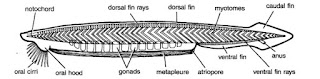


Protochordata:
These animals are bilaterally
symmetrical, triploblastic and have a
coelom. In addition, they show a new
feature of body design, namely a
notochord, at least at some stages during
their lives. The notochord is a long rod-like
supporting structure (chord=string) that
runs along the back of the animal separating
the nervous tissue from the gut. It provides
a place for muscles to attach for ease of
movement. Protochordates may not have a
proper notochord present at all stages in
their lives or for the entire length of the
animal.
Protochordates are marine animals.
Ex. Herdmania, Amphioxus.

 Herdmania


Amphioxus


BIO(Plasma Membrane)

                  Plasma Membrane


 Activity-1:

Get in-go out
Let us look at the substances in the
table. Some are used by the cells while
some are discarded by the cells.

Preparing Solutions:
To prepare sugar solution we need sugar
and water. In a sugar solution sugar is the
solute and water is the solvent. Sugar
dissolves in water forming sugar solution.

Preparation of saturated solution :
Take 100 ml of water in a beaker.
Add sugar/salt. Stir till it is dissolved.
Repeat it till little amount of it is left at
the bottom of the beaker which will not
dissolve. This is the saturated solution of
sugar/salt (in cold water).
Which one is more concentrated
solution?:
Take three beakers with one hundred ml.
of water in each. Add half teaspoon of sugar
to the first beaker, one teaspoon to the
second and one and a half teaspoon to the
third. Compare the three solutions and
answer the following. The solution of
which beaker will be most sugary? What is
the reason? Can we convert the solution of
beaker I into solution of beaker III?
How? How can we make the solution of the
third beaker indicated to that of the
first? How much water should we add to the
solution in the third beaker to make it
similar to solution of the first beaker?
Solutions with different amount of solute
dissolved in them are solutions of different
concentrations. The amount of sugar present
in the 100 ml of water is the concentration of the sugar.
Which beaker has the most concentrated solution?

Lab Activity:
Aim : Observation of material in different
solutions
Material: 1. Two beakers 2. Tap water
3. Sugar 4. Dry grapes or kishmish
Procedure: Take 100 ml of water in a
beaker. Keep dry raisin (kishmish) in it.


 Kishmish kept in tap water
Leave it for one hour. Observe what has
happened. Take it out and compare it with
the dry raisin. Is there any change in the
size of kishmish. (You may try the same
activity with slightly dried carrots and other
such vegetables) Did you ever observe that
your mother dipped wilted vegetables in
water. Can you identify the reason.
Then take 100 ml of saturated solution
of sugar in a beaker, which was already
prepared.

Swollen kishmish kept in tap water


Keep swollen kishmish of the previous
activity or fresh grape or carrot in it.
Leave it overnight. And observe in the
next morning what happens. Do you find
any change in the size of kishmish.
In the lab activity - 2a, water is
moving from …...............…… to
………….............
In the lab activity - 2b, water is moving
from …....……..… to ………......….
Let us think how water goes in and
comes out of Kishmish. Does the layer
permits water to pass through?. How does
it works? The cells in the outer layer of
kishmish permit water to pass through.
Learn more about the process. Let us
do the following activity.

Lab Activity:
Osmosis
Materials needed: One raw potato,
beaker / bowl, two pins, Water, Sharp knife.
Procedure: Take the raw potato and boiled
potato. Peel off the skins. Cut cube shaped
cups from them as shown in the figure (you
could make some other shapes as well).
Prepare a few amount of saturated sugar
solution.
Pour the sugar solution in the potato
cup to a level as shown in the figure.
Mark the level of sugar solution by
piercing the pin.
Keep the potato cup in the bowl/beaker

    scoop to make a cup


Pour water in the beaker / bowl to half
of the height of the potato cup. Ensure that
the potato cup does not float or submerge
in water.
Leave the arrangement for half an hour
and note your observations
Repeat the above experiment by taking
sugar solution in the beaker and water in
the potato cup. Note your observation and
compare with that of your previous one.
By comparing potato and kishmish
activities, do you find anything in common
among them? What is it?
From your observations you can
conclude that water always moved towards
the sugar solution. What might be the
reason? Such process, in which water moves
towards sugar solution is called Osmosis.
(In Greek Osmos means pushing).
We see that the movement of water
through membrane in osmosis i.e. from
less solute concentrated to more solute
concentration. To understand how materials may be
moving in and out of cells through the cell
membrane, let us do the following activity.


Activity-2:

Filtration


To perform this activity we need the
following materials:
Two beakers, Funnel, Filter paper,
Retort stand, Sugar, Iodine and Wheat / rice
flour, 500ml plastic bottle, cotton cloth.
Procedure :
1. Arrange the filtering apparatus as
shown in the figure (4a) or the
alternative method as in figure (4b).
2. Prepare wheat or rice powder
solution in a beaker by adding one
tea spoon of powder in 100ml of
water.
3. Add a drop of tincture iodine to the
solution.
4. Now pour the solution into the
funnel.
Then observe,
What remains on the filter paper or
the cloth?
What did the filter paper/ cloth
allow to pass through?
Which substance is not allowed by
the filter paper to pass through?
Why are certain substances not
allowed to pass through the filter
paper? Guess.
Cells also act in the same way while
allowing the substances to pass through the
plasma membrane.
With the understanding of above
activities let us try to understand the nature
of the Plasma membrane.
a) It allows water to pass through it.
b) It allows certain materials dissolved in
water to pass through.
c) It will not allow certain materials to
pass through it.
Allowing materials to pass through is
called permeability.
In the previous activities we have
observed the movement of water and
materials in plants.
To know more details about plasma
membrane or cell membrane, let us go
through the following paragraphs.
The outermost, extremely delicate,
elastic and membranous covering of the
cell that separates its contents from the
external environment is called plasma
membrane. Protoplasm of a cell consists
of two parts, cytoplasm and nucleus.
Plasma membrane is selectively
permeable because it allows entry of
certain substances, exit of some
substances while preventing passage to
remaining substances. Plasma membrane is
a living flexible membrane.

Functions of plasma membrane
Shape: It provides a definite shape to semi
fluid contents of the cell.
Mechanical Barrier: It functions as a
mechanical barrier that protects the internal
contents of the cell.
Selective Permeability: The membrane
determines what substances are to be
allowed entry or exit from the cell.
Endocytosis: The flexibility of the
membrane enables the cell to engulf food
and other substances (foreign particles)
from its external environment by
endocytosis. Amoeba acquires its food by
this process.
Recognition: It has substances over its
surface which function as recognition
centres and points of attachment. They help
in tissue formation, distinction of foreign
substances and defence against microbes.
Flow of Information :Helps for flow of
information amongst different cells of the
same organism.
Osmosis: It occurs due to presence of tiny
water channels in the plasma membrane.
Cell Continuity: At places plasma
membranes of adjacent cells become
continuous to form plasmodesmata and cell
junctions.
Specialization: Plasma membrane gets
modified to perform different functions,
e.g. absorption in microvilli.


Diffusion
There are other ways in which materials
move in a medium. Let us study one such
process by the following activity.
If a bottle of scent is opened in one
corner of a room, what will happen?
How do we feel? Its smell spreads in
the entire room. Let us think of the
following.
How does the smell spread in the
entire room?
Is the smell spreading uniformly in
the entire room?
Can you reach the scent bottle if
you are blind folded?
When blind folded how you are
able to locate the scent bottle?
Can you give any other such
examples?
There are other processes in which
substances move in a medium. Let us study
another such process by the following
activity.

Activity-4:
Diffusion with coffee powder
Experiment with coffee powder. Let us
Take half bowl of water. Prepare a small
ball of coffee powder. Slowly put it in
water. Observe what happens. Write a note
on what you observed.
Do this activity as many ways as you
could like.
Putting the pinch of coffee powder
first and adding water slowly.
Pouring hot water on the pinch of
coffee powder etc.
Note your observations for each situation
and write down what you infer.
Let us see whether other substances
also behave in the same manner.

Activity-5
Keep a small Crystal of KMnO4
(Potassium permanganate) in the
centre of the Petridish with the help
of a Forceps.
Carefully fill the Petridish with
water. (It is better to use filler or
dropper)Observe the movement of Pink
Colour in the Petri dish everyminute.
Also observe the spreading of colour
from center to periphery.
Repeat the experiment with other
substances (E.g.: copper sulphate) and
compare the result.
Do you find any relation between
Time and Movement?
For better understanding, the
movements of molecules in water, observe
the following activity.


BIO(Animal tissues)

             Animal tissues


 Activity-1:

Collect the substance lining the mouth
by using wooden spoon and observe this
under the microscope. Draw the diagram
that you have observed in the microscope,
in your note book.

How are the cells arranged?
Are there any inter cellular spaces?
The epithelial tissue, extremely thin
and flat, form a delicate lining. This is called
as squamous epithelium. We find this type
of epithelium in oesophagus, lining of
mouth, lining of blood vessels, lung alveoli
where transportation of substances
selectively occurs through permeable
membrane (you will learn about
permeability in the next chapter of
“Transport across Membrane)”.


The epithelial cells in skin are arranged
in the form of layers. This is called as
stratified squamous epithelium.
Think, why are the epithelial cells
in the skin are arranged in the form
of layers?
If you drink hot tea or chilled cool
drink, how do you feel?
If your skin burns or is wounded
which tissue would get effected?


Activity-2:
Take a permanent slide of cuboidal
epithelium from your laboratory slide box
and observe under the microscope. Draw
the picture in your note book. How are the
cells arranged?


These are the cuboidal epithelial cells
which form the lining of organs or tubules
like ureters or other parts and provide
mainly mechanical support to salivary glands.    

Activity-3:
Take a permanent slide of columnar epithelium from the slide box and observe it under the microscope.


Draw the figure that you have
observed under the microscope
How are the cells? Do you find any
hair like projections on the outer
surface of epithelial cells.
This types of cells are present where
ever absorption and secretion occurs. Try
to think where is this type of epithelial
tissues present in your body?
Do you know?The skin is also a kind of
epithelial tissue. Where does nails, and hair
grow from. The scales of fishes, reptiles
and feathers of birds also grow from
epithelium. These are modified epithelial
tissues. You learn more about them in the
chapter: Adaptations in different
Ecosystems.

Connective tissue:
If you tilt your body to any side of your body, what will happen to your internal organs? Is there any displacement? The internal organs are located at specific places without any displacement in organs
due to connective tissues. The tissueconnect organs and muscles. These tissuesare called connective tissue.


Connective tissues help in binding the
other tissues and organs together and
provide a frame work and support to various
organs in the body. These tissues also play
a major role in the transport of material
from one tissue to another. They also help
in the body defence, body repair and
storage of fat. There are different types of
connective tissues, each performing a
different function.

How do glasswear items carry for
longer distance?

Areolar tissue is one type of
connective tissue which joins different
tissues. It helps in packing and helps to keep
the organs in place. These cells are called
fibroblasts. These are the major
components in this type of connective
tissue. These cells secrete fibrous material
which holds the other tissue in position.
These cells also help in repair of the tissues
when they are injured.

           Areolar Tissue


The muscles in our body are attached
to the skin and bone by this type of tissue.
We can see this type of tissue around blood
vessels and nerves.


Why do old people shiver more in winter when than youngsters? Is there any special arrangement to prevent the escape of heat
energy during winter? Fat storing adipose tissue is found below the
skin and between internal organs. The cells of this tissue are filled with fat globules. Storage of fat also acts as insulator. Are all tissues in our body smooth and soft? Which tissue gives definite shape to
body of vertebrae? Bone is another type of connective tissue; it forms the frame work that supports the body. It is a major component of the skeletal system of several vertebrae
(except some fishes like sharks).


Cartilage is a type of connective tissue
found in the joints of bones, tip of ribs, tip
of the nose, external ears and in trachea.
Embryos of several vertebrae do not have
bone but have cartilage. The entire skeleton
of fishes like sharks is made of cartilage.
Cartilage is hard but not as hard as bone


How two bones are connected at joints?
Ligament is yet another type of
connective tissue that connects bones at
the joints and holds them in position. It is
made up of large number of fibres. These
fibres are made up of a protein called
collagen. This is very elastic in nature.

Activity-4:
Invite a scientist or doctor to your
place. Record an interview about blood
structure and its functions. It is important
to make a questionnaire inorder to conduct
interview. After completion of interview,
prepare a booklet about blood and keep it
in the class room library or display it on
the bulletin board.


It differs from other types of
connective tissues. There are different
types of cells in blood and each one has a
different function. All the cells in the blood
float freely in the plasma. Extra cellular
space is filled with fluid called plasma.
There are no fibres in blood.

Blood:
Blood is also a tissue which is having
different components. Let us know more
about blood.
There is a red stream that flows in
closed canals in our body. Think what is it?
The red stream that flows in closed tubes
in our body is blood. It is also a type of
connective tissue. Blood explains many
things about us. Blood is the source to
identify our wellness or illness. It is very
interesting to know about the blood current
in our body. There is highly sophisticated
and well developed mechanism to circulate
the blood to the entire body. Our heart pumps
nearly 36 thousand litres of blood to a
distance of nearly 20 thousand kilometers
in the time period of 24 hours. Blood is red
in colour. Do you agree with the statement
that all animals carrying red blood are your
blood relatives? The blood is always not red
in all animals. The cockroach has white
blood where as there is blue coloured blood
in snail. It is really a wonder that blood
appears in different colours.
Normal adult human beings have about
5 litres of blood. A chief component in
blood is water, which is stored in the
plasma.
Besides water, it also has several
nutrients such as glucose, amino acids,
proteins, vitamins, hormones, required for
the body and also excretory products such
as lactic acid, urea, salts etc. Plasma also
contains factors responsible for blood
clotting. Heparin helps to prevent blood
clotting in blood vessels.
Cells present in blood are corpuscles.
They are three types 1. RBC, 2. WBC,
3. Blood platelets.

Red blood cells also known as
erythrocytes which are red in colour. They
have red coloured protein called
haemoglobin. Because of haemoglobin
blood is red in colour, which helps in the
transport of oxygen and carbon dioxide 1ml
of human blood has about 5 millions of red
blood cells which live for 120 days in
blood.

Taking blood of an adult we can make a
chain of red blood cells around the earth at
equator with 7 circles. When you are in
your mother’s womb your RBC are formed
in the liver and spleen. After your birth
these RBC are generated from the bone
marrow of long bones. In mammals, mature
red blood cells are without nucleus.

Muscle Tissue:
If you are wounded deeply, a deep scar
would form at the place of wound? If we
are wounded on skin, a lighter scar would
form. Why? For the reason, that the skin
cells have regenerating character. Think
about the muscle cell. Will they get
regenerated like epithelial cells?
Muscles are responsible for the
movements of hands and legs and also of
several internal organs such as intestine and
heart. Small amounts of muscle tissues also
present in blood vessels. These helps in
increasing or decreasing the diameter of
the blood vessel and thus the blood flows.
Heart is made of only one type muscle cells
and they help in pumping the blood.

         Striated muscles


     Non-Striated muscles


How do muscles contract and relax?
Muscular tissue consists of elongated
cells called muscle fibres. This tissue is
responsible for movement in our body.
Muscles contain special protein called
contractile proteins which contract and
relax to cause movement.
During winter, body shivers. Why?
When the body is exposed to cold air,
we shiver. During shivering muscles
contract and relax producing large amount
of heat. This keeps the body hot.
Based on their structure, location and
function, muscles are three types. They are
striated muscle, non-striated muscle,
cardiac muscle.
We can move some muscles by our
conscious effort. For example the muscle
present in inner limbs move according to
our wish and will Such muscles are called
voluntary muscle. These muscles also
called as skeletal muscles as they are
mostly attached to bones and help in body
movement; these muscles show alternate
light and dark bands or striations. As a result,
they are also called striated muscle. The
cells of this tissue are long, cylindrical, un
branched and have many nuclei in the
cytoplasm (multi nucleated).


Nervous Tissue:
If you put your fingers in a glass of hot
water, how do you feel?
How would you know the water is hot
or cold? If you put your leg on a sharp edged
stone while walking, how will you feel?
The feelings like the above situations
is because of specialized mechanism in our
body. It works like electric current passing
through wires. Brain, spinal cord and nerves
play active role in this mechanism.

            Nerve cell


Cell body or cyton has a large nucleus
and cytoplasm. The cytoplasm contains
granular structure called Nissal’s granules.
There are some projections arising
from cell body. These are called dendrite.
They are sharp, branched, more in number.
One projection of the cyton is somewhat
longer than remaining projections.
This is called axon. Some nerve cells have axon
covered with sheath like structure. This
sheath is called as Myeline sheath. Gas or
nodes present at regular intervals on
myeline sheath are known as Ranvier
Nodes.

Axon of a nerve cell is connected with
Dentrites of a near by nerve cell to form a
web like structure throughout bod


BIO(Plant tissues)

              Plant Tissues


         Plants that we observe around us are
usually multi-cellular. They perform
several life processes such as growth,
respiration, excretion, etc, similar to those
performed by animals. In addition to these
they can perform photosynthesis and
prepare food not only for themselves but
also for all the other living organisms dependent on them, either directly or indirectly. Let us recall the information about different parts of the plants and the functions they are associated with.

Activity-1:
Parts of the plant and their
functions.
We have studied about the functions of
the parts of the plant in the earlier classes.
Read the functions given below and write
the names of the parts involved in
performing the respective function.

    Function Name of the parts:
1. Absorption of water from soil
2. Exchange of gases (air)
3. Photosynthesis
4. Transpiration
5. Reproduction

Activity-2:
Cells in onion peel
- Take a piece of an onion peel.
- Now place it on the slide.
- Put a drop of water and then a drop of
glycerine on it.


- Gently cover it with a cover -slip.
- Observe it under the microscope.
- Draw and label the diagram that you
have observed under the microscope.
Compare your drawing with figure-1 to
find out labelled parts.
Are all the cells similar?
How are the cells arranged?

Activity-3:
Cells in a leaf peel.
- Take a betel leaf or Tradescantia or
Rheo leaf.
- Tear it with a single stroke. So that a
thin edge be seen at the torn end.
- Observe the thin edge where the leaf


has been torn under the microscope
in the same manner as you had
observed the onion peel.
Draw a diagram of what you have
observed and compare it with figure-2.
Are all the cells similar?
Is there any difference in their
arrangement?
What can we infer from the above
activities?
Have you noticed that the cells are in
groups in both the activities?
Compare and write a note on the
arrangements of the cells that you have
observed in both of the activities.
You may have observed that the cells are
present in groups with certain
arrangement. With the help of the
following activities we shall try to find out
whether these arrangements have special
roles to play in the plant body.

Activity-4:
(a) Cells in root tip
- Are the cells in the root similar to that
of a leaf. Let us find out how the cells
are arranged in the root. For this we
need onion root tips.
- Take a transparent (plastic/glass) bottle
filled with water. Take an onion bulb
slightly larger than the mouth of the
bottle.
- Put the onion bulb on the mouth of the
bottle.


- Observe the growth of roots for a few
days till they grow to nearly an inch.
- Take the onion out and cut some of the
root tips.
- Take an onion root tip.
- Place it on the slide.
- Put a drop of water and then a drop of
glycerine on it.
- Cover it with a cover-slip.
- Remove excess water with the help of
blotting paper.
- Tap the cover-slip gently and press with
the blunt end of the needle or brush to
spread the material.
- Observe the structure and arrangement
of the cells.
- Draw the diagram of that you have
observed under the microscope.
· Are all the cells similar?
· How is the arrangement of cells?


       Cells in Onion roots tip:


Activity-5:
(b) Growing roots
- Take the onion used in the previous
activity and mark the cut end of the
roots with a permanent marker.
- Put the onion bulb on the mouth of the
bottle in the same way as you did in the
previous activity.
- Leave the set up aside for at least four
to five days. Take care that there is
enough water in the glass so that the
roots are submerged.
Did all the roots grow in a same manner?
What happened to the roots which had
been cut off?
Write down your findings, regarding the
cut roots and those that were not?
We observe that by removing the tip of
the onion root, having a particular
arrangement of cells, the growth of the root
in length is stopped.
You have observed that cells are present
in groups. Cells in groups which are nearly
similar in structure perform similar
functions. Such groups of cells are called tissues.


      Growth on roots in onion bulb

Types of plant tissues:
There are four basic types of tissues in
the plants. They are
Tissues that bring about overall growth
and repair are called Meristematic
tissues.
Tissues that form outer coverings are
called as Dermal tissues.
Tissues that form the bulk of the plant
body, helping in packing other tissues
are called as Ground tissues.
 Tissues that help in transport of
materials are called as Vascular tissues.
You have already observed some types
of tissues. To observe various types of plant
tissues we need to know some techniques
for preparing slides and cutting sections.
See annexure for the same.

1. Meristematic tissues:
Observe the given figure of a stained
section of a shoot tip


From the above figure you can infer
that meristematic tissues are present on
the tip, sides and in between layers of other
tissues. Meristematic tissues at the
growing tip that bring about growth in
length are called as apical meristematic
tissues.
Tissues present around the edges in a
lateral manner and giving rise to growth in
the girth of stem are called lateral
meristematic tissues.
Areas from which branching takes place
or a leaf or a flower stalk grows, we find
a kind of meristematic tissue called
intercalary meristematic (also called
Cambium) tissue.


Activity-6:
Dicot Stem tissues
Prepare a temporary mount of the TS
of a dicot stem.
Observe it under the microscope.
Draw and label the diagram.
Compare it with figure given below

       Dicot stem (TS) - Tissues


What are the similarities between the
tissues in root and in shoot as shown
in the above figure?
Are all the cells similar in shape and
structure?

Activity-7:
Rheo leaf - Dermal tissue. 
- Take a fresh leaf of Rheo or Betel plant
- Tear it in a single stroke, so that a thin whitish edge can be seen at the torn end.
Slowly remove it and observe that peel
under the microscope (by preparing a
temporary mount).
See the arrangement of cells .Are all
cells similar? Are there any spaces
between the cells?
This activity shows a part of the dermal
tissue of the plants.
Dermal tissue (Dermis) usually consists
of a single layer of cells showing variations
in the types of cells on the basis of their
functions and location. These tissues are
divided into three different types. They are
epidermis (outer most layer), mesodermis
(The middle layer) and endodermis (the
innermost layer).
The walls of the cells of dermal tissue
are comparatively thicker than the cells of
meristematic tissues. In desert plants it may
be even more thick and waxy. Small pores
are seen in the epidermis of the leaf, called
stomata. They are enclosed by two kidney
shaped cells, called guard cells. Cells of
the roots have long-hair like parts, called
root hair.


        Parenchyma:
The cells of the parenchyma are soft,
thin walled and loosely packed. The
Parenchyma which contains chloroplasts is
called “Chlorenchyma”. The Parenchyma
which contains large air cavities or spaces
is called “Aerenchyma”. The Parenchyma
which stores water or food or waste
products is called “Storage Tissue”.
Collenchyma tissues have thick walled,
longer cells when compared with parenchyma. In the sclerenchyma the cells are thick walled and tightly packed without spaces. So when compared with dermal tissue, the ground tissue is of various types. And some of the ground tissues help in photosynthesis. Let us observe the ground tissue of some other stems.


Activity-8:
Take permanent slides of Chlorenchyma, Arenchyma, Storage Tissue from your laboratory and observe them under the microscope. Find out the characteristics differences and record them in your notebook.

       Chlorenchyma


             Arenchyma


          Storage Tissue


      Vascular Tissue:
We know that roots can absorb water
from the soil and send it to other parts of
the plant. The leaves and other green parts
prepare food and supply it to all the parts
of the plant.
Let us study the tissues involved in
transportation.
We have done an experiment on
transportation in class VII, in the chapter
on plant nutrition. We have observed that
when a plant is kept in red coloured water,
Some of the parts of the plant turned red.
Do the same experiment again by keeping
a small plant (with roots) in red coloured
water. Leave it for two hours. Now cut a
T.S. of the stem and observe it under the
microscope.
Which part of the plant is responsible
for this transport?
Draw a rough sketch of that part and
mark the part that appears red.
What do you conclude from your observation?
The tissues involved in transportation are vascular tissues. They are composed of different
types of cells which show specific arrangements.